Ear
ANATOMY
In plain talk, the auditory system is comprised of three components; The External, Middle, and Inner ear, all of which work together to transfer sounds from the environment to the Brain.
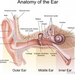
The External Ear
It includes the portion of the ear that we see—the pinna/auricle and the ear canal.
Pinna
The pinna or auricle is a concave cartilaginous structure, which collects and directs sound waves through air into the ear canal or external auditory meatus.
Ear Canal
The ear canal or external auditory meatus is approximately 1.25 inches long and .25 inch in diameter. The inner two-third of the ear canal is imbedded in the temporal bone. The external one-third of the canal is cartilage. Although the shape of each ear canal varies, in general the canal forms an elongated “s” shape curve. The ear canal directs airborne sound waves towards the tympanic membrane. The ear canal resonate sound waves and increases the loudness of the tones in the 3000-4000 Hz range.
The ear canal maintains the proper conditions of temperature and humidity necessary to preserve the elasticity of the tympanic membrane. Glands, which produce cerumen (ear wax) and tiny hairs in the ear canal, provide added protection against insects and foreign particles from damaging the tympanic membrane.
Middle Ear
The middle ear is composed of the tympanic membrane and the cavity, which houses the ossicular chain.
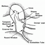
Tympanic Membrane
The tympanic membrane or eardrum serves as a divider between the external ear and the middle ear structures. It is gray-pink in color when healthy and consists of three very thin layers of living tissue.
The eardrum is very sensitive to sound waves and vibrates back and forth as the sound waves strike it. The eardrum transmits the airborne vibrations from the external to the middle ear and also assists in the protection of the delicate structures of the middle ear cavity and inner ear.
Middle Ear Cavity
The middle ear cavity is in the mastoid process of the temporal bone. The middle ear cavity extends from the tympanic membrane to the inner ear. It is approximately two cubic centimeters in volume and is lined with mucous membrane.
The middle ear cavity is actually an extension of the naso-pharynx via the Eustachian tube.
Eustachian Tube
The Eustachian tube acts as an air pressure equalizer and ventilates the middle ear. Normally the tube is closed but opens while chewing or swallowing. When the Eustachian tube opens, the air pressure between the external and middle ear is equalized. The transmission of sound through the eardrum is optimal when the air pressure is equalized between the external and middle ear. When the air pressure between the external and middle ear is unequal, the eardrum is forced outward or inward causing discomfort and the ability of the eardrum to transmit sound is reduced.
Ossicular Chain
The middle ear is connected and transmits sound to the inner ear via the ossicular chain. This chain consists of the three smallest bones in the body: the malleus, incus, and stapes. The malleus is attached to the tympanic membrane. The footplate of the stapes inserts into the oval window of the inner ear. The incus is between the malleus and the stapes.
Attached to the ossicular chain are two tiny muscles, the stapedius and tensor tympani muscles. These muscles contract to protect the inner ear by reducing the intensity of sound transmission to the inner ear from external sounds and vocal transmission.
The ossicular chain amplifies a signal approximately 25 decibels as it transfers signals from the tympanic membrane to the inner ear.
The Inner Ear
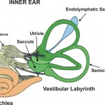
The inner ear is composed of the sensory organ for hearing—the cochlea, as well as for balance—the vestibular system. The systems are separate, yet both are encased in the same bony capsule and share the same fluid systems.
Vestibular or Balance System
The balance part of the ear is referred to as the vestibular apparatus. It is composed, in part, of three semicircular canals located within the inner ear. The vestibular system helps to maintain balance, regardless of head position or gravity, in conjunction with eye movement and somatosensory input. The semicircular canals are innervated by the VIIIth cranial nerve.
Cochlea
The hearing part of the inner ear is the cochlea. The cochlea is spiral-shaped, similar to the shape of a snail.
The cochlea is composed of three fluid-filled chambers that extend the length of the structure. The two external chambers are filled with a fluid called perilymph. Perilymph acts as a cushioning agent for the delicate structures that occupy the center chamber. It is important to note that perilymph is connected to the cerebrospinal fluid that surrounds the brain and the spinal column. The third fluid filled chamber is the center chamber, called the cochlear duct. The cochlear duct secretes a fluid called endolymph, which fills this chamber.
The cochlear duct contains the Basilar membrane upon which lies the Organ of Corti. This Organ of Corti is a sensory organ essential to hearing. It consists of approximately 30,000 finger-like projections of cilia that are arranged in rows. These cilia are referred to as hair cells. Each hair cell is connected to a nerve fiber that relays various impulses to the cochlear branch of the VIIIth cranial nerve or auditory nerve. The “pitch” of the impulse relayed is dependent upon which areas of the basilar membrane, and hence, which portions of the Organ of Corti are stimulated. The apical portion of the basilar membrane (the most curled area of the cochlea) transfers lower frequency impulses. The basal end relays higher frequency impulses.
The VIII cranial nerve (VIII C.N.) or auditory C.N. carries the impulses generated from the Organ of Corti to the brainstem. From the brainstem, nerve pathways extend through numerous nuclei to the cerebral cortex in the temporal lobes of the brain. It is in the temporal lobes of the brain that meaning is associated with the various patterns of nerve impulses.
THE PHYSIOLOGY OF HEARING
The process of hearing begins with the occurrence of a sound. Sound is initiated when an event moves and causes a motion or vibration in air. When this air movement stimulates the ear, a sound is heard.
In the human ear, a sound wave is transmitted through four separate mediums along the auditory system before a sound is perceived: in the external ear—air, in the middle ear— mechanical, in the inner ear liquid and to the brain—neural.
Sound Transmission through the External Ear
Air transmitted sound waves are directed toward the delicate hearing mechanisms with the help of the external ear, first by the pinna, which gently funnels sound waves into the ear canal, then by the ear canal.
Sound Transmission through the Middle Ear
When air movement strikes the tympanic membrane, the tympanic membrane or eardrum moves. At this point, the energy generated through a sound wave is transferred from a medium of air to that which is solid in the middle ear. The ossicular chain of the middle ear connects to the eardrum via the malleus, so that any motion of the eardrum sets the three little bones of the ossicular chain into motion.
Sound Transmission through the Inner Ear
The ossicular chain transfers energy from a solid medium to the fluid medium of the inner ear via the stapes. The stapes is attached to the oval window. Movement of the oval window creates motion in the cochlear fluid and along the Basilar membrane. Motion along the basilar membrane excites frequency specific areas of the Organ of Corti, which in turn stimulates a series of nerve endings.
Sound Transmission to the Brain
With the initiation of the nerve impulses, another change in medium occurs: from fluid to neural. Nerve impulses are relayed through the VIII C.N., through various nuclei along the auditory pathway to areas to the brain. It is the brain that interprets the neural impulses and creates a thought, picture, or other recognized symbol.